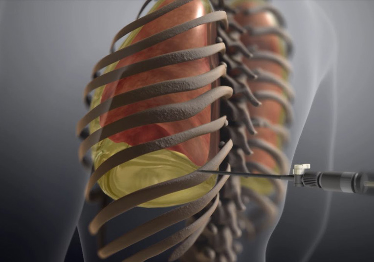
Thoracentesis

Thoracentesis is a procedure to remove fluid from the space between the lungs and the chest wall, or the pleural space. Thoracentesis involves a needle camera (and sometimes a plastic catheter) being inserted through the chest wall. Ultrasound pictures are often used to guide the placement of the needle. Once the fluid has been removed from the pleural space, it may then be sent to a lab to determine what is causing the fluid to build up in the pleural space.
Depending on the volume of fluid in the pleural space, this procedure generally takes 10 – 15 minutes to complete.
Although a thoracentesis usually does not cause serious problems, some risks are have been noted. Common risks include pneumothorax (collapsed lung), pain, bleeding, bruising, or infection at the spot where the needle or tube was inserted.


